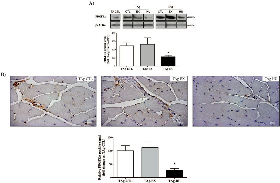
Fig. 3. Effect of different muscle activity levels on expression of FAP cell surface marker PDGFRα. A) PDGFRα protein levels from TAg of each experimental group (CTL, EX and HU), 21 days after glycerol muscle injury (N=6). B) Representative PDGFRα immunostained histological transversal paraffin-embedded from TAg of each experimental group (CTL, EX and HU), 21 days after glycerol muscle injury and quantification of the PDGFRα-positive signals (N=4). One-way ANOVA was used to compare our experimental groups,* p<0.05 vs. TAg-CTL.